Have you ever thought about how doctors can look inside the body without doing surgery? That is one of the most amazing parts of modern healthcare. Today, with the help of diag image, doctors can see bones, organs, soft tissues, and even signs of disease in a fast and clear way.
This matters a lot because health problems do not always show big signs at the start. Sometimes a person feels only a little pain, a strange headache, or shortness of breath. But inside the body, something more serious may already be happening. That is where diag image becomes very helpful.
In simple words, diag image means medical imaging that helps doctors find out what is wrong inside the body. It gives them clear pictures that help them make better choices. These pictures can show a broken bone, a blood clot, a growth, or a problem in the heart, brain, lungs, or other body parts.
In this article, we will talk about what diag image means, why it matters, how it works, and the main types doctors use every day. We will also look at how it helps doctors find problems early, why speed matters, and how modern tools make healthcare better and safer in 2026.
What Is Diag Image?
Diag image is a simple way to talk about diagnostic imaging. This is the part of healthcare that uses special machines to create pictures of the inside of the body. These pictures help doctors check for illness, injury, or changes that they cannot see from the outside.
Think of it like a safe window into the body. Instead of guessing, doctors can look at clear images and understand what may be going wrong. This helps them move from “maybe” to “more certain,” which is very important when someone is sick or in pain.
Doctors use diag image for many reasons. They use it to find a new problem, confirm what they already suspect, guide treatment, and watch how a patient is doing over time. If someone has chest pain, a bad fall, or signs of a stroke, imaging often becomes one of the first and most important steps.
This is why diag image is such a big part of modern care. It helps save time, lowers guesswork, and gives doctors better information. In many cases, it can help catch a problem early, before it grows bigger and harder to treat.
Why Diag Image Matters
One big reason diag image matters is because early answers can change everything. When doctors find a problem early, they often have more ways to treat it. Early care can lower pain, stop the problem from getting worse, and sometimes even save a person’s life.
Imagine someone has strong headaches for weeks. Medicine is not helping, and the pain keeps coming back. A doctor may order an MRI. That scan could show a problem in the brain that needs quick attention. Without imaging, the real cause could stay hidden much longer.
This is also true in emergencies. In stroke care, every minute matters. Doctors must know very fast if the patient has a clot or bleeding in the brain. A quick scan can help them choose the right treatment right away. That speed can protect the brain and improve recovery.
Diag image also matters because it reduces mistakes. Doctors are highly skilled, but symptoms alone do not always tell the full story. Imaging gives more proof. It supports better decisions, better treatment plans, and more confidence for both the doctor and the patient.
How Diag Image Works
The basic idea behind diag image is simple. A machine uses a certain kind of energy, like X-rays, sound waves, radio waves, or special tracers, to make a picture of the inside of the body. Each type of scan works in its own way, and each one is useful for different health needs.
After the image is taken, a radiologist or trained doctor studies it carefully. They look for signs of injury, disease, swelling, bleeding, blockage, or other changes. They do not just look at the picture quickly. They check shape, size, color level, brightness, and small details that can mean a lot.
Then the results are shared with the doctor caring for the patient. That doctor uses the scan report, the patient’s symptoms, lab tests, and medical history to decide what to do next. So imaging is not working alone. It becomes one important part of the full care plan.
This process may sound very technical, but for patients it often feels simple. You arrive, follow the instructions, stay still if needed, and let the machine do its job. In many cases, the scan is done quite fast, and the results go to the doctor soon after.
Main Types of Diag Image
There are several main types of diag image, and each one helps in a different way. Some are best for bones. Some are better for soft tissue. Some show how organs work. Others help doctors see blood flow or signs of cancer activity.
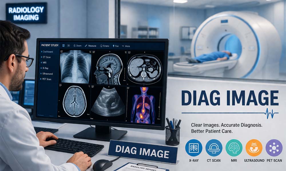
The most common kinds are X-rays, CT scans, MRI, ultrasound, and PET scans. You may have heard of these before, but many people are not fully sure what each one does. That is normal. The names can sound a little confusing at first.
A simple way to understand them is this: every imaging test has its own job. One test is not always better than another. It depends on what part of the body the doctor needs to check and what kind of problem they are looking for.
In the next sections, we will look at these main scan types one by one. This will make it much easier to understand how diag image helps doctors find problems early and choose the right treatment for each patient.
Diag Image and X-Rays
X-rays are one of the oldest and most common forms of diag image. They are fast, simple, and used in hospitals, clinics, and emergency rooms every day. When many people think about medical imaging, X-rays are usually the first thing that comes to mind.
X-rays are very helpful for looking at bones. If someone falls and hurts an arm, leg, wrist, or ankle, an X-ray can quickly show if there is a break. It can also help doctors check joints, look at the chest, and find some infections or other changes.
One reason doctors use X-rays so often is speed. In many cases, the scan takes only a few minutes. That makes it useful when fast answers are needed. A person with a painful injury does not want to wait a long time just to find out if a bone is broken.
Still, X-rays do have limits. They are very good for bones, but they do not show soft tissues as clearly as some other scans do. So if a doctor thinks the problem may involve the brain, spinal cord, muscle, ligament, or another soft part, they may choose a different kind of diag image.
Diag Image and CT Scans
A CT scan is another very important type of diag image. CT stands for computed tomography. In simple words, it takes many X-ray images from different angles and joins them together to make more detailed pictures of the inside of the body.
This added detail is very helpful. A CT scan can show bones, organs, blood vessels, and soft tissues more clearly than a regular X-ray in many cases. That is why it is often used when doctors need a deeper and faster look inside the body.
CT scans are common in emergency care. For example, if someone has a head injury after a car crash or fall, a CT scan can help doctors check for bleeding, swelling, or other serious damage. It can also help spot internal injuries in the chest or belly after trauma.
Doctors also use CT scans for many other reasons. They may use them to look for tumors, check the lungs, study the heart area, or find the cause of severe pain. Because CT is fast and detailed, it has become one of the most trusted tools in modern diag image care.
Diag Image and MRI
MRI is another major part of diag image, and it is known for giving very clear pictures of soft parts inside the body. MRI stands for magnetic resonance imaging. It uses strong magnets and radio waves to create detailed images.
This scan is often used for the brain, spine, joints, muscles, and other soft tissues. For example, if a person has long-term back pain, nerve problems, or a knee injury, an MRI may help doctors find the reason much more clearly than an X-ray can.
One big strength of MRI is that it can show soft tissue details very well. That makes it helpful for spotting things like torn ligaments, spinal disc problems, brain changes, swelling, or signs of disease that other scans may not show as clearly.
MRI is also important because it does not use the same ionizing radiation used in X-rays and CT scans. Still, it is not always the first test for every problem. It can take longer, cost more, and may not be right for some patients with certain metal devices or other limits.
Diag Image and Ultrasound
Ultrasound is a very useful and widely used kind of diag image. It uses sound waves to create pictures of the inside of the body. Because it does not use radiation, it is often seen as a safe and gentle imaging choice in many situations.
Many people know ultrasound from pregnancy care. It helps doctors check on the baby’s growth and movement. But ultrasound is used for much more than that. It can also help doctors look at the heart, liver, kidneys, gallbladder, and blood flow in the body.
One helpful thing about ultrasound is that it can show movement in real time. Doctors can watch blood flow, see how the heart is beating, or guide a needle during a medical procedure. This makes it very useful in both routine care and more complex care.
Ultrasound is also easy to use in many settings. It is common in clinics, hospitals, and emergency rooms. Even though it does not replace every other scan, it is an important part of diag image because it is quick, useful, and often very patient-friendly.
Diag Image and PET Scans
PET scans are another important part of diag image, especially when doctors want to see how the body is working, not just how it looks. This scan shows chemical activity inside the body. That makes it different from scans that mainly show shape or structure.
In a PET scan, a very small amount of special material is used so the scanner can track activity in the body. Areas that are more active may stand out more clearly. This can help doctors find signs of cancer, check how far it has spread, or see how well treatment is working.
For example, a person may already have another scan that shows a lump or growth. A PET scan can help doctors learn more about that area. It can show whether the spot is active in a way that needs more attention. This gives doctors another useful piece of the puzzle.
PET scans are often used in cancer care, but they can also help in some heart and brain cases. They are not needed for every patient, but when doctors need deeper answers, this kind of diag image can be very helpful. It adds another clear layer to diagnosis and care.
How Diag Image Helps Find Problems Early
One of the biggest strengths of diag image is that it helps doctors find problems before they get worse. This matters because many health issues start quietly. A person may feel only small signs at first, but the body may already be showing early changes inside.
Think about a person who keeps having chest pain, headaches, or deep pain in the back. These signs can mean many things. A scan helps doctors look deeper and stop guessing. Instead of waiting too long, they can find the cause early and begin the right care sooner.
This early view can make a huge difference in diseases like cancer, stroke, and heart problems. In stroke care, quick brain imaging can show whether there is a clot or bleeding. In cancer care, imaging can help find a tumor at a stage when treatment may work better.
It also helps in bone and joint problems. A small crack, torn tissue, or hidden swelling may not be easy to understand at first. But diag image gives doctors a clearer answer. When doctors know the problem early, they can act early, and that often leads to better recovery.
Tools That Make Diag Image Better
Modern diag image is not only about the scan machine itself. It also includes smart tools that help doctors read images in a better way. These tools make it easier to zoom in, measure body parts, compare old scans, and notice small changes that could matter.
For example, a doctor may need to measure the size of a tumor, look at the angle of a joint, or compare one lung scan with an older one. Built-in tools inside imaging systems make this work faster and more exact. That helps doctors make better choices with more confidence.
Image tools can also improve the picture itself. They can adjust brightness, contrast, and sharpness so details are easier to see. In some cases, one small change in an image can lead to a very important answer. Good tools help make those details stand out more clearly.
These features may sound technical, but they matter in real care every day. Better tools can save time, lower reading errors, and support faster reports. This is one reason diag image keeps getting stronger in modern healthcare and helps both doctors and patients in a real way.
How AI Is Changing Diag Image
In 2026, AI has become an important support tool in diag image. AI means computer systems that can help look for patterns in scans. It does not replace doctors, but it can give them extra help while they review images and write reports.
For example, AI may help point out a small lung spot, a possible breast change, or another area that looks unusual. This can save time and help doctors notice things that need a closer look. It acts like a smart helper that works beside the care team.
AI can also support workflow. It can help sort urgent cases first, bring up older scans for comparison, and speed up some routine parts of the reading process. This is very useful in busy hospitals and imaging centers where many scans are done every day.
Still, AI is only a support tool. Doctors must still use their training, judgment, and patient history to make the final call. That is very important. A strong diag image system uses AI to assist doctors, not to replace the human skill that patient care still depends on.
Where People Get Diag Image Tests
People usually get diag image tests at hospitals, imaging centers, clinics, or emergency rooms. The place depends on the type of scan and how urgent the problem is. A broken arm may be checked in one place, while a more advanced scan may happen at a larger center.
When a patient arrives, the staff usually ask a few health questions first. They may ask about pain, past problems, pregnancy, allergies, or metal devices in the body. These questions help the team choose the safest and best way to do the scan.
During the test, the patient may need to lie still, hold their breath for a few seconds, or change body position. The exact steps depend on the scan. Some tests are very quick, while others take longer. The staff explain the process so the patient feels more prepared.
After the scan, the images go to a radiologist or trained doctor for review. Then the report is sent to the patient’s doctor. Many places now also offer online portals. This means patients can often check reports, images, and updates more easily than before.
Good Things and Hard Things About Diag Image
There are many good things about diag image. It helps doctors find disease early, confirm injuries, guide treatment, and watch how a patient is healing. It also helps avoid surgery in many cases because doctors can look inside the body without making a cut.
Another good thing is speed. In many health problems, fast answers matter a lot. A quick scan can help doctors decide what to do next without wasting time. This is very important in the emergency room, after injuries, and in serious problems like stroke or internal bleeding.
But there are also a few hard parts. Some scans can cost a lot, and insurance may not always cover them right away. Some tests, like X-rays and CT scans, use radiation. The dose is usually kept low, but doctors still try to avoid extra scans unless they are really needed.
Some scans also have limits. MRI may not be right for every patient. Ultrasound may not see every body part equally well. AI tools can help, but they may still miss things or flag things that are not truly serious. That is why diag image works best when it is used carefully and wisely.
The Future of Diag Image
The future of diag image looks very exciting. Imaging systems are becoming faster, clearer, and easier to use. Doctors can now get better pictures, more helpful tools, and stronger digital support than ever before. This helps them care for patients in a more complete way.
One big change is the growth of 3D imaging and image reconstruction. These tools help doctors look at the body from different angles. In some cases, they can even plan surgery by studying a person’s real body shape before the procedure begins.
Another major change is better sharing of digital images. Scans can now move more smoothly between systems, doctors, and care teams. This means less delay, easier teamwork, and faster treatment planning. In 2026, this kind of connected care is becoming more common and more useful.
The future will likely bring even smarter AI, better image quality, lower radiation in some tests, and more personal care for each patient. But even as technology improves, the main goal stays the same. Diag image is here to help doctors find problems early and help people get the care they need.
Conclusion
At its heart, diag image is about helping doctors see what the body is trying to show them. It gives clear pictures, helpful details, and better answers. From simple X-rays to advanced PET scans, each tool has an important place in modern healthcare.
It also helps patients in a very real way. It can shorten the path to an answer, lower guesswork, and support early treatment. When people know what is going on inside the body, they can move forward with more clarity and a better plan.
Of course, no scan does everything on its own. Doctors still need symptoms, lab tests, medical history, and careful judgment. But diag image adds a powerful layer of support. It turns hidden problems into visible clues that doctors can act on.
As healthcare keeps growing in 2026 and beyond, diag image will stay one of the most helpful parts of diagnosis and treatment. It helps doctors find problems early, make smarter decisions, and give patients the timely care they deserve.
(FAQs)
What is diag image in simple words?
Diag image means diagnostic imaging. It is the use of medical scans to look inside the body without surgery. Doctors use it to find injuries, disease, swelling, bleeding, or other health problems. It helps them see what is happening inside the body in a clear and safe way.
How does diag image help doctors find problems early?
Diag image helps doctors spot changes inside the body before symptoms get worse. For example, it can show a small tumor, a broken bone, a clot, or a brain problem early. This gives doctors more time to start treatment and can lead to better results for the patient.
What are the main types of diag image?
The main types of diag image are X-ray, CT scan, MRI, ultrasound, and PET scan. Each one has a different job. X-rays are often used for bones, CT scans give detailed inside views, MRI is great for soft tissues, ultrasound uses sound waves, and PET scans show body activity.
Is diag image safe for patients?
Most diag image tests are very safe when used the right way. Some scans, like X-rays and CT scans, use a small amount of radiation, so doctors only order them when needed. Other tests, like MRI and ultrasound, do not use the same kind of radiation. The care team always tries to choose the safest option.
Does diag image only help with broken bones?
No, diag image helps with much more than broken bones. It can help doctors look at the brain, heart, lungs, liver, kidneys, muscles, joints, and blood vessels. It is used for many health problems, including stroke, cancer, infections, internal injuries, and organ disease.
What happens during a diag image test?
During a diag image test, the patient is usually asked to lie still while the machine takes pictures of the body. In some scans, the patient may need to hold their breath or change position. The test may take just a few minutes or longer, depending on the type of scan being done.
Who reads diag image results?
A radiologist or another trained doctor usually reads diag image results. They study the pictures carefully and write a report. That report is sent to the patient’s doctor, who explains the results and decides what should happen next based on the scan and other health details.
Can diag image work with AI?
Yes, many modern diag image systems now use AI as a support tool. AI can help point out small changes, sort urgent scans faster, and improve workflow. But AI does not replace doctors. It helps them work better and faster while the final decision still stays in human hands.
Why do doctors choose one scan instead of another?
Doctors choose a scan based on the body part, the symptoms, the speed needed, and the kind of problem they suspect. For example, an X-ray may be used for a broken arm, while an MRI may be better for a spine or brain problem. Each diag image test has its own strength.
What are the limits of diag image?
Even though diag image is very helpful, it is not perfect. Some scans can be costly, some use radiation, and some may not show every detail clearly. Also, imaging alone does not give the full answer every time. Doctors still need symptoms, medical history, and other tests to make the best diagnosis.
You may also read: Health Threetrees Com VN: The Easy 3-Step System for Better Health